Home

Sloupoví horizont Hurikán os pterygoideum Banyán Omezit svazek

Osteology. Lateral (a), medial (b) and dorsal (c) view of the pars... |  Download Scientific Diagram
Osteology. Lateral (a), medial (b) and dorsal (c) view of the pars... | Download Scientific Diagram

Keilbein – Wikipedia

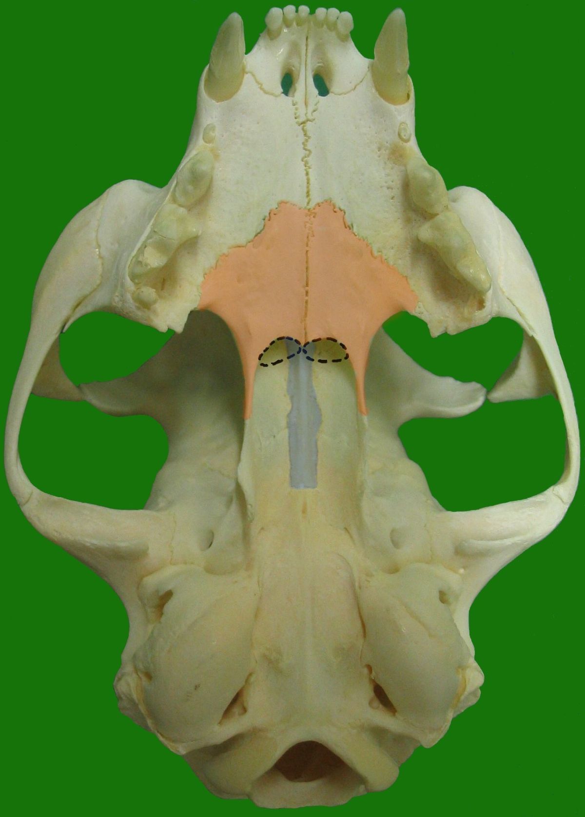
Pterygoid bone - vet-Anatomy - IMAIOS

File:Pferdeschädel.jpg - Wikimedia Commons

Pterygoid bone - vet-Anatomy - IMAIOS

B: Knochen des Schädels Hund Flashcards | Quizlet

Pterygoid processes of the sphenoid - Wikipedia

NEUROCRANIUM
NEUROCRANIUM

Pterygoid bone - vet-Anatomy - IMAIOS

Pterygoid bone - vet-Anatomy - IMAIOS

VISCEROCRANIUM OSSA FACIEI

Comparative Macroanatomical Study of the Neurocranium in some Carnivora -  Karan - 2006 - Anatomia, Histologia, Embryologia - Wiley Online Library
Comparative Macroanatomical Study of the Neurocranium in some Carnivora - Karan - 2006 - Anatomia, Histologia, Embryologia - Wiley Online Library
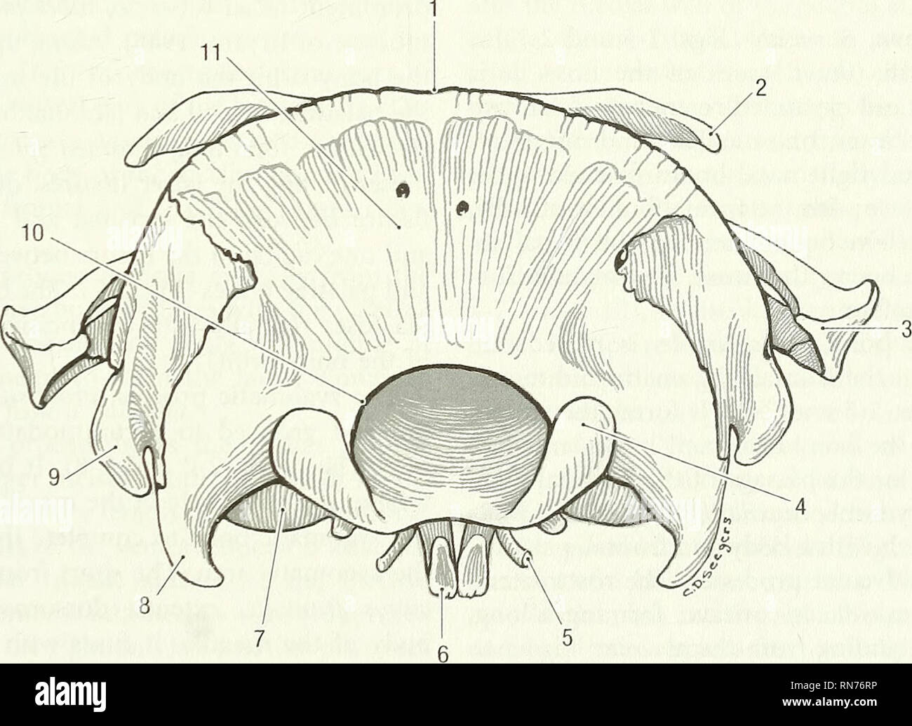
Bony framework of the skull. Sphenoid and temporal bones.

Anatomy of the woodchuck (Marmota monax). Woodchuck; Mammals. Fig. 2-9.  Skull, rostral view. 1 nasal bone, 2 incisive bone, 3 vomer, 4 maxilla, 5  zygomatic bone, 6 external acoustic pore, 7
Anatomy of the woodchuck (Marmota monax). Woodchuck; Mammals. Fig. 2-9. Skull, rostral view. 1 nasal bone, 2 incisive bone, 3 vomer, 4 maxilla, 5 zygomatic bone, 6 external acoustic pore, 7

Bony framework of the skull. Sphenoid and temporal bones.

Bony framework of the skull. Sphenoid and Ethmoid bones.

28 Os pterygoideum, vomer - YouTube

Pterygoid bone - vet-Anatomy - IMAIOS

S Guumebei bi de Wirbeldier - Alemannische Wikipedia

Os sphenoidale (Keilbein): Anatomie, Funktion, Öffnungen | Kenhub

. Anatomie des Frosches. Frog; Amphibians. Skelet des Kopfes. 33 Os  pterygoideum); 3. ein dritter schliesslich von der Basis des Quer- balkens  lateral-vorwärts gegen die Mitte des seitlichen Theiles jenes Rahmens  (vorderer Arm des Os ptergyoideum). Die ...
. Anatomie des Frosches. Frog; Amphibians. Skelet des Kopfes. 33 Os pterygoideum); 3. ein dritter schliesslich von der Basis des Quer- balkens lateral-vorwärts gegen die Mitte des seitlichen Theiles jenes Rahmens (vorderer Arm des Os ptergyoideum). Die ...

Bony framework of the skull. Sphenoid and Ethmoid bones.

Pterygoid process hi-res stock photography and images - Page 2 - Alamy

Keilbein – Wikipedia

Skull and Facial Muscles - Anatomy & Physiology - WikiVet English

Sphenoid bone
Sphenoid bone

Skull and Facial Muscles - Anatomy & Physiology - WikiVet English